


Pathology detection platform
The pathology detection platform has strong comprehensiveness and professionalism. In terms of histopathology, it can accurately process samples and produce high-quality slices, clearly presenting the microstructure of tissues. Immunohistochemistry testing is equipped with abundant antibody resources, and precise technology is used to locate and analyze intracellular antigens in tissues, providing key information for tumor biomarker analysis, antibody screening, and cytokine detection. Pathological diagnosis is provided by a senior team based on tissue pathology and immunohistochemistry results, combined with clinical data to provide accurate conclusions and report interpretation consultation. To provide users with comprehensive and high-quality pathological support, laying a solid foundation for accurate diagnosis and in-depth research in the future.Our services
Experiment Name | content | describe |
Pathological section | Decalcification of bone tissue | Slow release, fast release |
Paraffin embedding | Fixed sample | |
OCT embedding | Fresh/Fixed Samples | |
paraffin section | Fixed sample | |
frozen section | Fresh/Fixed Samples | |
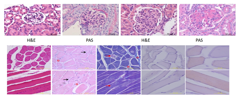
Pathological staining | HE dye |
|
Pathological staining | Masson Trichrome、Oil red O、Sirius Red、 PAS、IHC、Multiplex Immunofluorescent、 Masson-Fontana、Gorky staining | |
TUNELApoptosis Detection | bright field | |
immunohistochemistry | General immunohistochemistry | Single antibody |
Indirect fluorescence method | Multi label (3 labels and 4 colors) | |
Construction of immunofluorescence TSA fluorescence method scheme | Multi label (3 labels and 4 colors) | |
Pathological imaging | Full slice scanning (bright field) -20X/40X |
|
Immunofluorescence whole section scanning-20X | Single label and multi label | |
Pathological morphological description |
Case sharing