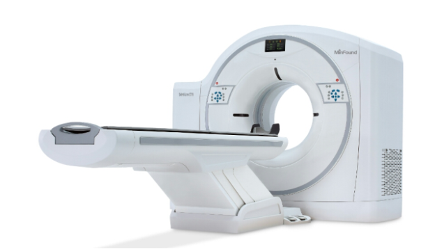
Large and small animal CT
CT is a non-destructive 3D imaging technology that can clearly understand the internal microstructure of the sample without destroying the sample. CT uses X-ray beams to scan the size of a certain part of an animal with a certain thickness. The detector receives the X-rays transmitted through the beam and converts them into visible light. The electrical signals are converted by photoelectric conversion, and then the analog/digital converter (analog /Digital converter) to convert into numbers, input into the computer for processing. The image formation process is like having several cuboids with the same volume on each side, called voxels.The scanned information is calculated to obtain the X-ray attenuation coefficient or absorption coefficient of each voxel, and then arranged into a matrix, that is, a digital matrix. The digital matrix can be stored in a magnetic disk or an optical disc. The digital/analog converter converts each number in the digital matrix into small squares with varying gray levels from black to white, namely pixels, and arranges them in a matrix to form a CT image. Therefore, CT images are reconstructed images. The X-ray absorption coefficient of each voxel can be calculated by different mathematical methods.
Application areas
Central nervous system disease diagnosisDiagnosis of head and neck diseases
Diagnosis of chest disease
CT examination of abdominal and pelvic diseases
Case sharing


