


Do you know that over 80% of the wrinkles, pigmentation, and sagging on our faces are caused by "photoaging" - the long-term "kissing" of the sun's ultraviolet rays. In order to fight against time, scientists first need to "recreate aging" in the laboratory, and mice and guinea pigs have become the first "ray chasers".
Today, let's take you into the laboratory to see how these magical animal models can help us uncover the secrets of skin aging.
1. Why them?
The unique application of the three major star models
Not all animals are suitable for photoaging research. Scientists have carefully considered the choice between mice and guinea pigs.
Ⅰ. Common Applications: Basic Mechanisms and Drug Screening
Both mice and guinea pigs can be used for studying the mechanism of UV induced skin aging, such as exploring the roles of oxidative stress, inflammatory response, collagen degradation (such as MMPs matrix metalloproteinase activation), DNA damage repair, and other pathways in photoaging; At the same time, it is also a core model for screening anti-aging drugs/skincare products - by comparing the skin appearance, pathological structure, and molecular indicators of the intervention group and the model group, the anti-aging effect of the product is evaluated (such as whether it can reduce wrinkles and increase collagen content).
Ⅱ. Differentiated Applications: Species Characteristics Determine Research Directions
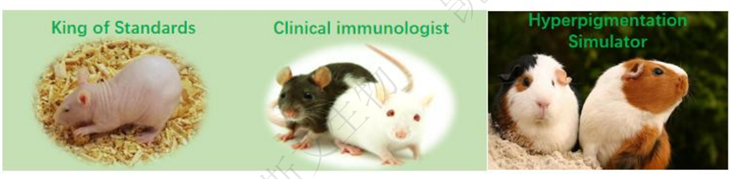
• Hairless Mice: The King of Standardized Research
Advantages: Hairless, avoiding the obstruction of UV rays by hair, allowing UV rays to act evenly and efficiently on the skin, resulting in more consistent and highly reproducible experimental results.
Application: It is a gold standard model for studying the mechanism of photoaging (such as collagen degradation, oxidative stress) and screening anti photoaging drugs/cosmetics. Most of the well-known anti-aging ingredients you can think of have been validated on it.
• Hairy mice (C57BL/6, etc.): a special talent for studying "immunity" and "hair follicles"
Advantages: Although hair needs to be shaved, its genetic background is clear and the immune system is thoroughly studied.
Application: Very suitable for studying how ultraviolet radiation affects the skin immune system (such as suppressing immunity and increasing the risk of skin cancer) and the role of hair follicle stem cells in the process of photoaging.
• Guinea Pig: The Best Stand in for "Color Depth" Research
Advantages: Its skin structure and melanin metabolism are very similar to those of humans! It is easier to simulate pigmentation (sunspots) caused by human exposure to ultraviolet radiation.
Application: It is an ideal model for evaluating whitening and spot lightening products. Want to verify if a certain ingredient can remove spots? First, ask the guinea pig for their opinion.
2. How to 'create' aging?
Core methods for establishing photoaging models
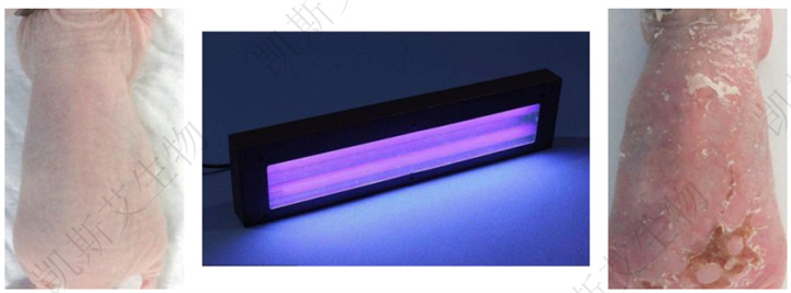
Sunbathing in the laboratory is not that simple, it is a precisely controlled science.
Ⅰ. Light source selection:
In natural ultraviolet radiation, UVA (320-400nm) and UVB (280-320nm) are the main bands that cause photoaging:
UVB mainly damages the epidermis, causing DNA damage (such as cyclobutane pyrimidine dimer formation) and inflammatory reactions; UVA penetrates deeper and mainly causes degradation of dermal collagen and oxidative stress. Therefore, modeling commonly uses "UVA+UVB mixed irradiation" or single band irradiation (depending on the research objective):
• If simulating overall photoaging: choose a mixed light source of UVA (dose ratio of 70% -80%) and UVB (dose ratio of 20% -30%), which is closer to the natural UV effect.
• If focusing on epidermal damage: use UVB irradiation alone; If focusing on dermal aging (such as wrinkles, collagen loss), use UVA irradiation alone.
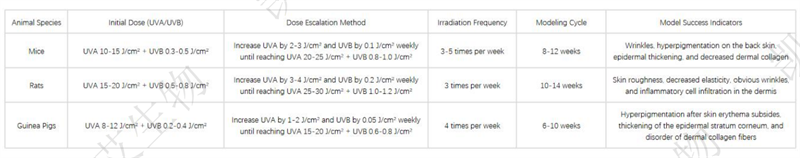
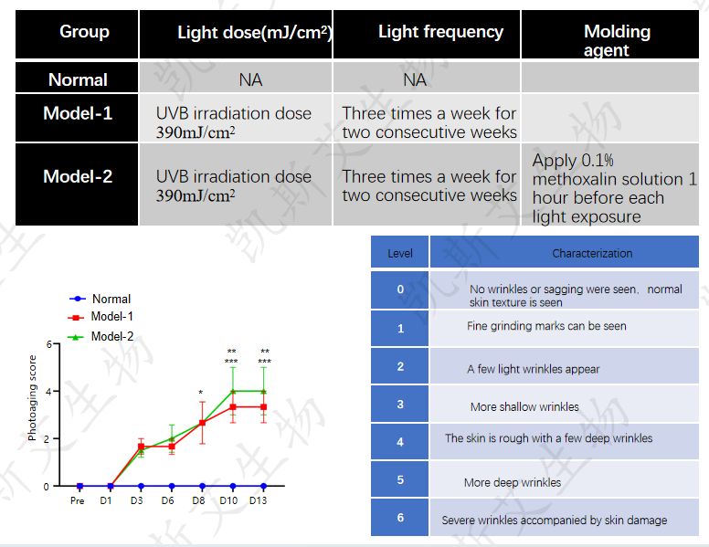
Ⅱ. Exposure plan: gradual and avoid acute injury
The core principle is to gradually increase the dose, allowing the animal skin to adapt to the irradiation and avoiding acute burns (such as blisters and ulcers) caused by excessive dose, which may affect the stability of the model. The classic parameter reference is as follows:
Ⅲ. Modeling precautions: Reduce interference factors
• After shedding hair, hairy mice need to wait for 24 hours before starting irradiation to restore the skin barrier;
• Confirm the stability of the light source power before each irradiation (calibrated with a UV radiometer);
• During irradiation, fix the position of the animal and ensure that the irradiation area (such as the 2cm x 3cm range on the back) is precise, avoiding exposure to other parts of the body;
• During the modeling period, observe the animal's diet and weight. If severe skin damage occurs, the irradiation should be suspended.
3. The evidence is conclusive!
From 'macro' to 'micro' validation model
The success of the photoaging model needs to be comprehensively judged through "appearance observation+histopathological examination" - appearance reflects the overall phenotype, while pathological sections reveal the microscopic changes in each layer of the skin, which is the gold standard for model validation.
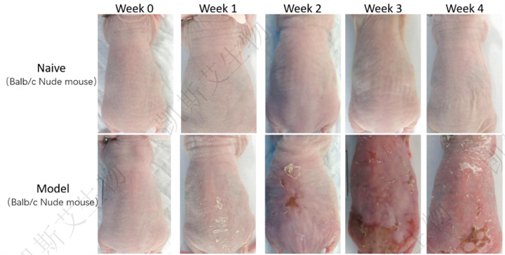
Ⅰ. Macroscopic appearance evaluation: intuitive judgment of aging phenotype
After the modeling is completed, observe the following indicators with the naked eye or skin detector:
• Wrinkles: Use a skin wrinkle analyzer to capture images of the back skin and analyze the number and depth of wrinkles (wrinkles are more pronounced in mice and guinea pigs, and relatively shallow in rats due to thicker skin);
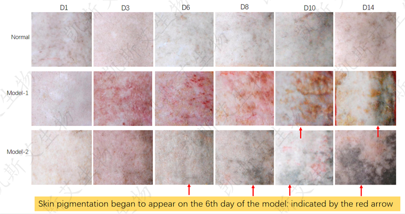
• Pigmentation: Observe changes in skin color (for example, guinea pigs are prone to obvious pigmentation, and mice with black skin need to compare differences in pigmentation depth);
• Barrier function: The integrity of the skin barrier is detected using a transcutaneous water flow analyzer (TEWL), and the photoaging model usually shows an increase in TEWL values (barrier damage).
Ⅱ. Histopathological examination: dismantling changes in various layers of the skin
(1) Sample preparation: standardized sampling and slicing
• Sampling: After the modeling is completed, the animals are euthanized, and the skin of the back irradiation area (2cm × 1cm) is taken to remove subcutaneous fat, which is fixed with 4% paraformaldehyde for 24 hours;
• Slicing: After dehydration and paraffin embedding, 4 μ m thick continuous slices are made for HE staining (observing tissue structure), Masson trichrome staining (observing collagen fibers), and immunohistochemical staining (detecting specific molecules).
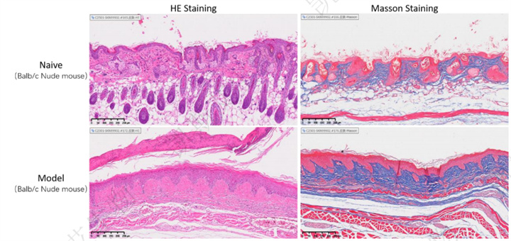
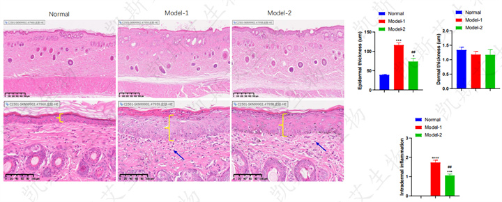
(2)HE staining: Observing the structure of epidermis and dermis
Typical changes in HE staining of photoaging model:
• Epidermal: Thickening of the epidermis (especially the stratum corneum and stratum spinosum), disordered cell arrangement, and basal cell proliferation (compensatory proliferation after UVB induced epidermal injury);
• dermis: increase or decrease in thickness of the dermis layer (UVA mainly causes collagen degradation in the dermis, and thickness may decrease); UVB induced inflammation, with possible increase in thickness, and infiltration of inflammatory cells (such as neutrophils and lymphocytes);
• Other: Some models show dermal capillary dilation (more pronounced in guinea pigs).
(3) Masson trichrome staining: evaluation of collagen fibers
Collagen fibers are the main structural component of the dermis, and photoaging can lead to collagen degradation and disorder. In Masson staining, collagen fibers appear blue and can be evaluated by the following indicators:
• Proportion of collagen fiber area: Use analysis software such as Visioparma to analyze the proportion of the blue area in the section to the dermis area. The photoaging model usually shows a decrease in proportion (collagen loss);
• Collagen arrangement: Normal skin collagen fibers are arranged neatly and parallel to the epidermis; The photoaging model showed collagen fiber breakage, disorder, and disordered arrangement (collagen changes were more significant in mice and guinea pigs, while collagen in rats was thicker and the changes were relatively less significant).
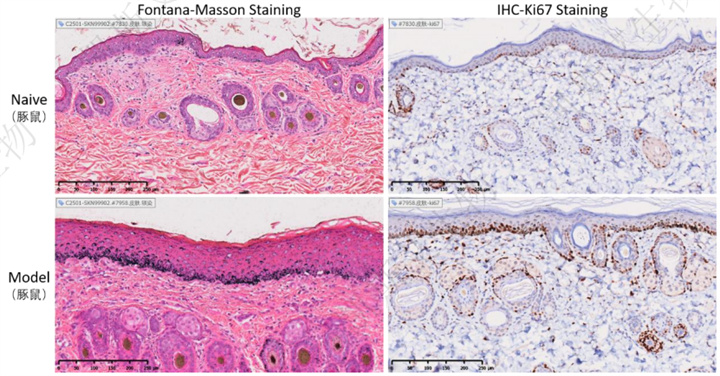
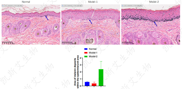
(4) Fontana Masson staining: evaluation of melanin deposition
The melanin in normal guinea pig skin is mainly distributed in the basal layer of the epidermis (cytoplasm of melanocytes), and its content is relatively low; After long-term exposure to ultraviolet radiation (UV, such as UVA+UVB), the photoaging model guinea pigs exhibit "light induced pigmentation" similar to that of humans. Fontana Masson staining can clearly show an increase and aggregation of melanin particles throughout the epidermis (especially in the basal and spinous layers), even extending to the stratum corneum or the superficial dermis where free melanin particles appear (abnormal migration of melanocytes).
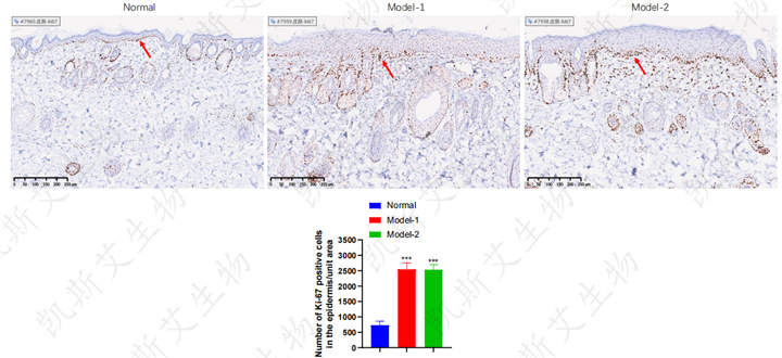
(5) Immunohistochemistry/Immunofluorescence: Detection of Specific Molecular Markers
Select biomarkers based on research objectives to validate the activation of photoaging related pathways:
• Cell proliferation: Detecting Ki67 can accurately evaluate changes in the proliferation activity of skin cells. In normal skin, Ki67 positive cells are mainly concentrated in the basal layer of the epidermis (with only a few in the proliferative phase, maintaining epidermal renewal); In the photoaging model, UV irradiation significantly increases the number and distribution range of Ki67 positive cells (extending from the basal layer to the spinous layer or even the granular layer), indicating "abnormal proliferation" of the epidermis (compensating for UV induced cell damage or apoptosis).
• DNA damage: detection of gamma H2AX (DNA double strand break marker) or Cyclobutane Pyrimidine Dimers (CPD, UVB induced DNA damage marker), with increased positive expression in epidermal cells of photoaging models;
• Collagen degradation: Detection of MMP-1 (matrix metalloproteinase 1, a key enzyme in collagen degradation), and an increase in MMP-1 positive cells in the dermis of the photoaging model;
• Inflammatory response: Detection of TNF - α (tumor necrosis factor alpha) or IL-6 (interleukin-6), with increased positive expression in dermal inflammatory cells in a photoaging model.
4. KCI Biotech Skin Disease Pharmacology and Pharmacology Evaluation Platform
UVB induced photoaging animal (guinea pig) model
Experimental animals:
• Brownish A1 Guinea guinea pig, male, 5-6w
Testing indicators:
• General observations
• Skin observation: before modeling, before each subsequent irradiation after modeling
• Skin rating: before modeling, before each subsequent irradiation after modeling
End point anatomy:
• serum/plasma
• Back skin tissue
• histopathology of skin
▪ Gross observation of the skin
▪ Pathological analysis of skin tissue: HE
▪ Pathological analysis of skin tissue: IHC-Ki-67
▪ Pathological analysis of skin tissue: melanin staining
5. Summary
Selecting appropriate models to assist in photoaging research
The photoaging models of mice and guinea pigs each have their own advantages: mice and rats are suitable for gene mechanisms and high-throughput screening, while guinea pigs are more closely related to human skin phenotypes; The core of model establishment is "precise control of ultraviolet parameters", while pathological diagnosis requires a combination of macroscopic appearance and microscopic structure. Regardless of which model is chosen, it is necessary to make a comprehensive judgment based on research objectives (such as mechanism exploration, product screening), experimental period, cost, and other factors to ensure the stability and practicality of the model.
In the future, with the development of gene editing technology (such as CRISPR) and imaging technology (such as laser confocal microscopy), photoaging models will more accurately simulate human skin aging, providing more powerful tools for anti-aging research.