Introduction
In the microscopic world of life science research, how can we accurately track the in vivo transport of substances and localize the distribution of target molecules? Fluorescent labeling technology serves as a "luminescent key" to solve this problem. Among relevant techniques, the combination of in vivo injection of fluorescent tracers and subsequent histological examination plays an irreplaceable role in disease mechanism research, drug development, cell behavior tracing and other fields.
Logical Framework of This Scientific Sharing
1 Why Choose In Vivo Injection of Fluorescent Tracers?Fluorescent labeling technology conjugates fluorescent substances with target molecules or carriers. After administration into living organisms, the dynamic distribution of these substances can be tracked via specialized detection equipment. Compared with other labeling methods, it possesses unique advantages:
·Excellent visualization: Fluorescent signals are distinct and distinguishable, directly displaying the localization of tracers in tissues and cells, and even enabling real-time in vivo imaging.
·High specificity: Labeling with specific antibodies or aptamers allows precise targeting of target molecules and minimizes non-specific interference.
·Broad compatibility: Applicable to a wide range of experimental organisms (mice, rats, Drosophila, etc.) and labeling substrates (proteins, nucleic acids, drug carriers, etc.), with flexible application scenarios.
Combined with subsequent histological examination, further in-depth analysis of the interactions between tracers and tissue cells can be realized, providing solid morphological evidence for research conclusions.
2 Selection of Fluorescent Tracers
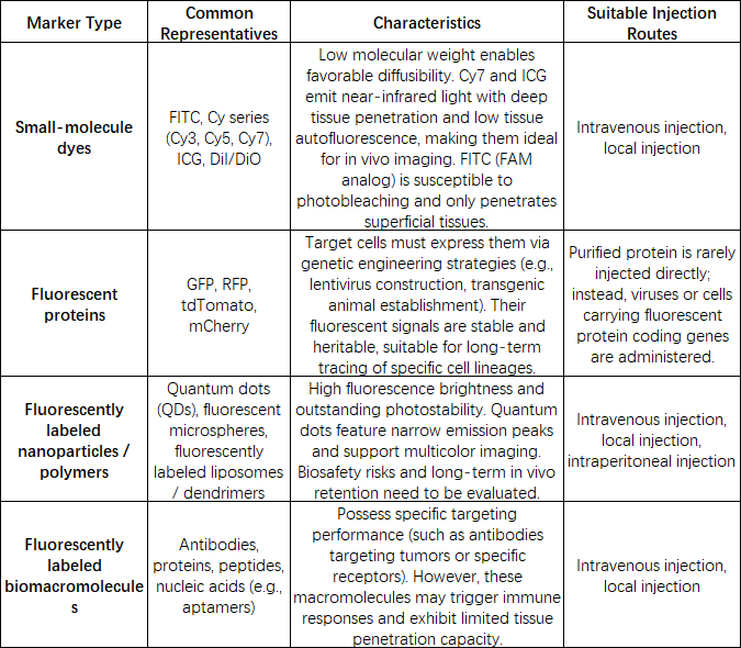
3 Key Points for In Vivo Injection of Fluorescent Tracers
Standardized injection procedures directly determine the reliability of experimental results. Every step from reagent preparation to practical operation must be strictly controlled.
1.Pre-injection Preparation:
·Selection of tracers: Fluorescent tracers for in vivo administration shall be selected based on research objectives, imaging penetration depth, signal duration and other factors. Candidates should feature favorable biocompatibility, low toxicity, stable fluorescence and strong tissue penetration to guarantee labeling efficiency and signal stability.
·Disinfection of instruments: Syringes and needles (33 Gauge is commonly used) shall be disassembled and autoclaved at 115 °C to prevent microbial contamination that may compromise experimental outcomes.
·Preprocessing of experimental animals: Appropriate anesthetics shall be selected according to animal species and injection sites (e.g., intraperitoneal injection of ketamine-xylazine mixture for mice). Anesthetic depth shall be assessed via toe pinch test after anesthesia. Hair covering injection sites shall be removed in advance; moisturizing eye drops shall be prepared for fragile ocular tissues to avoid dryness-induced damage.
2.Common Injection Routes & Operational Tips
Different injection sites are adopted for distinct research purposes. Core protocols for three widely used routes are listed below:
·Intravitreal injection: Mainly applied in ophthalmic research, requiring two operators. Secure the mouse laterally under a surgical microscope, gently pull the eyeball out of the orbital fossa with fine forceps, and puncture along the limbus (junction of cornea and sclera). Retract the needle slightly to drain a small volume of vitreous humor before slow infusion of tracer solution. Slowly withdraw the needle after injection and apply moisturizing eye drops.
·Tail vein injection: Suitable for systemic distribution studies. Select the well-vascularized middle segment of the mouse tail, dilate blood vessels with alcohol swabs, insert the needle at a 30° angle against the tail, and push the solution slowly. Blanching of the vessel confirms successful injection.
·Local tissue injection: For tumor tissue, muscle tissue and other localized regions. Precisely locate the target tissue, insert the needle vertically into the tissue parenchyma and inject slowly to prevent tracer leakage into adjacent normal tissues.
3.Post-injection Precautions
Place animals on a heating pad for recovery after injection, and continuously monitor their respiration and motor activity. Return animals to breeding cages only after complete wakefulness. Record injection time, dosage, animal status and other data to determine sampling time points for subsequent histological examination (e.g., tissues can be harvested 3 hours after intravitreal injection to detect tracer uptake).
4 Workflow of Histological Examination for Fluorescent Tracers
Tissue samples are collected at predetermined time points post injection, followed by fixation, sectioning, staining and imaging for histological analysis. The core of this procedure is to preserve intact tissue morphology while retaining fluorescent signals.
1.Tissue Harvest and Fixation:
Euthanize animals and rapidly dissect target tissues (retina, tumor, liver, etc.) to avoid hypoxic necrosis. Fixation is a critical step; appropriate fixation methods shall be chosen according to tracer properties:
·No fixation: Embed fresh tissues in OCT compound and snap-freeze on dry ice to prepare frozen tissue blocks.
·Fix with 4% paraformaldehyde at room temperature for approximately 6 hours: Ideal for preserving tissue morphology and antigen epitopes when detecting intranuclear antigens. Conduct gradient sucrose dehydration before OCT embedding for frozen block preparation.
·Fix with 4% paraformaldehyde at room temperature for approximately 12 hours: Perform gradient ethanol dehydration followed by paraffin embedding to generate FFPE (formalin-fixed paraffin-embedded) tissue blocks.
2.Sectioning:Sectioning protocols vary by tissue block type
Frozen sectioning: Cut sections at a thickness of 5–20 μm with a cryostat, mount sections onto glass slides and air-dry at room temperature.
Paraffin sectioning: Cut sections at a thickness of 3–10 μm with a microtome, mount sections onto slides, bake at 60 °C for 30 minutes, then conduct deparaffinization and hydration.
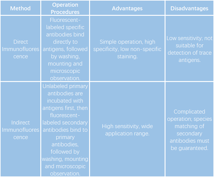
3.Staining:Fluorescent signals may attenuate during labeling or tissue processing, and counterstaining can be performed to amplify signals.
Two staining approaches (direct method and indirect method) are available for selection based on experimental requirements.
4.Mounting:
Mount slides with anti-fade mounting medium to mitigate light-induced photobleaching during observation and extend signal retention time.
5.Imaging & Result Interpretation:
Image samples under appropriate microscopes (fluorescence microscope, confocal laser scanning microscope), and select matching detection channels according to the emission spectrum of fluorescent tracers to avoid spectral crosstalk.
Key notes for result interpretation:① Exclude tissue autofluorescence via negative control groups (tissues from non-injected animals);② Restrict co-localization analysis to positively labeled or successfully transfected cell regions;③ Faint target fluorescence in unstained sections indicates signal attenuation, and counterstaining is recommended for re-observation;④ If only DAPI blue signal is detectable with weak target fluorescence in stained sections, insufficient antibody concentration or mismatched secondary antibody species may be the cause, and experimental conditions need optimization.
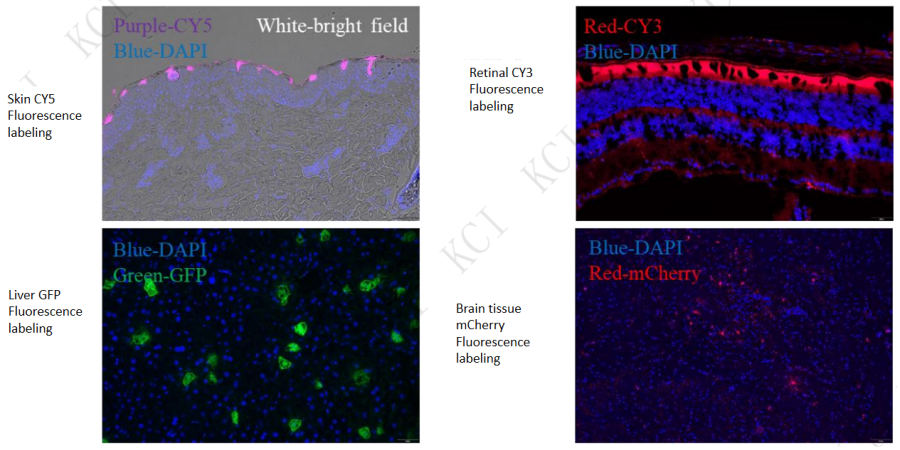
Case Sharing of In Vivo Injection of Fluorescent Markers and Histological Examination
KCI・KMQ Pathology Platform
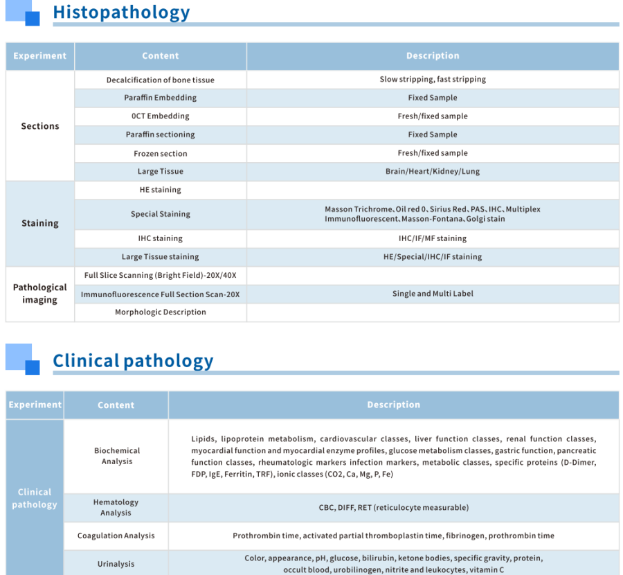
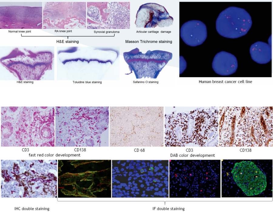
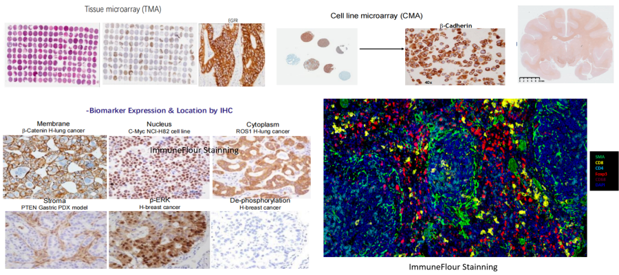
KCI・KMQ owns a comprehensive and powerful pathology platform, including histopathology platform, immunohistochemistry/immunofluorescence platform, clinical pathology platform and other sub-platforms. It delivers full-spectrum pathology services covering pathological anatomy, histopathology, immunopathology, digital pathology, tumor pathology, toxicological pathology and more. The technical team holds internationally recognized professional expertise, provides customized services for complex histological projects, and fully meets diverse detection demands.