Introduction
Reduced glutathione (GSH) is one of the most important intracellular antioxidants, and also serves as a critical biomarker for evaluating redox status, metabolic health and drug sensitivity. As laboratories have growing demands for highly sensitive and reproducible detection assays, a standardized and complete practical protocol for GSH detection becomes essential. Accurate GSH measurement provides key evidence for judging cellular redox state, assessing the antioxidant capacity of organisms, exploring the pathogenesis of diseases, and verifying the efficacy of drug intervention.This paper summarizes a full practical workflow for reduced glutathione (GSH) detection. The whole procedure, including sample pretreatment, reagent preparation, experimental operation, result calculation and troubleshooting of common problems, is broken down in detail for easy understanding, to help researchers perform GSH detection efficiently and accurately.

Experimental Principle
Reduced glutathione (GSH) reacts with 5,5'-dithio-bis-(2-nitrobenzoic acid) (DTNB) to produce thionitrobenzoic acid and glutathione disulfide. Thionitrobenzoic acid is a yellow compound, and the content of reduced glutathione (GSH) can be quantitatively determined by colorimetry at 412 nm. Detection range: 2–600 μmol/L, sensitivity: 2 μmol/L.
Main Instruments and Reagents
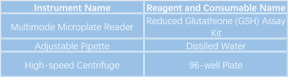

Multimode Microplate Reader
Experimental Procedures
1.Take out the kit in advance and equilibrate it to room temperature.2.Preparation of standard solution:Dissolve the standard with 1 mL of distilled water immediately before use to prepare a 10 mmol/L standard stock solution, which can be stored at 2–8 °C for 6 weeks. Take 200 μL of the 10 mmol/L standard solution and mix with 1800 μL of distilled water to obtain a 1 mmol/L standard mother solution. Dilute the 1 mmol/L standard mother solution with distilled water to prepare standard samples with the following concentration gradients: 0 μmol/L, 50 μmol/L, 100 μmol/L, 150 μmol/L, 200 μmol/L, 250 μmol/L, 300 μmol/L, 400 μmol/L.
3.Preheat the microplate reader for more than 30 min and set the wavelength to 412 nm.
4.Preparation of sample supernatant:Take 80 μL of the test sample, add 80 μL of Reagent 1 and mix well. Centrifuge at 4500 g for 10 min, then collect the supernatant for detection. If the supernatant contains precipitates, transfer it to a new EP tube and centrifuge again.
5.Sample determination (add reagents sequentially in a 96-well plate).
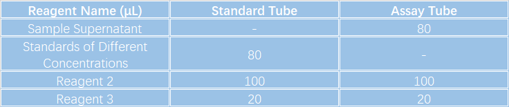
☑Mix thoroughly, incubate at room temperature for 5 min, then measure the absorbance of each well at 412 nm respectively.
6.Calculation of Experimental Results.Standard calibration curve fitting: \(y=ax+b\).
(y: OD value of standard minus OD value of blank well; x: concentration of standard; a: slope of standard curve; b: intercept of standard curve)
Notes
1. Do not use expired products. Components from different kits shall not be mixed.2. It is recommended to select 2–3 samples with widely different expected concentrations for a pre-test before the formal experiment. Samples can be properly diluted or concentrated according to the pre-test results.
3. Wear lab coats and latex gloves for protection during the experiment.
4. Store reagents strictly under specified conditions. Reagents from different test kits cannot be mixed. For reagents with small volumes, centrifuge the vial before use to ensure sufficient reagent can be pipetted.
KCI・KMQ Molecular Biology Platform
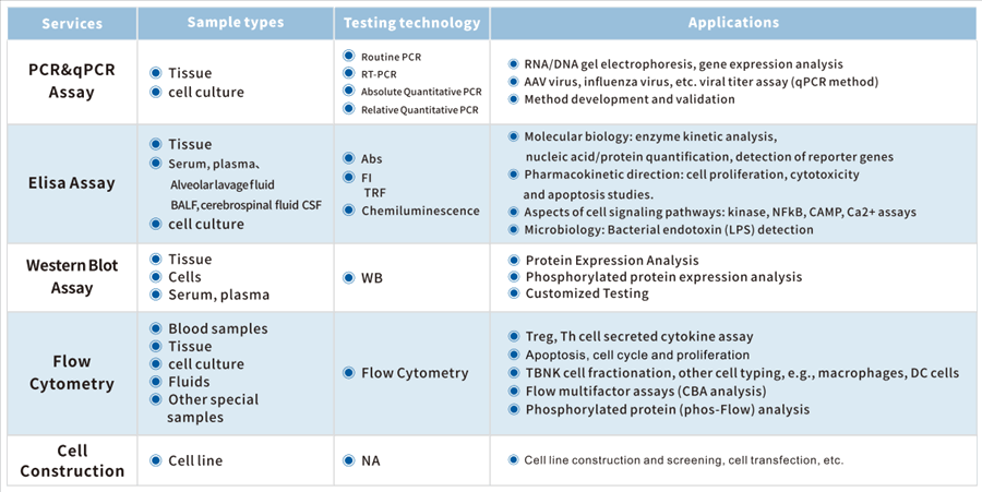
Equipped with advanced technologies and rich project experience, the KCI・KMQ Molecular Biology Platform can perform more than 300 biochemical detection assays per day. It also provides a full spectrum of molecular biology detection services including flow cytometry analysis, qPCR, ELISA, WB and IP/Co-IP.Up to now, the company has established extensive long-term partnerships with numerous well-known pharmaceutical enterprises and research institutions at home and abroad, laying a solid foundation for innovative drug development.